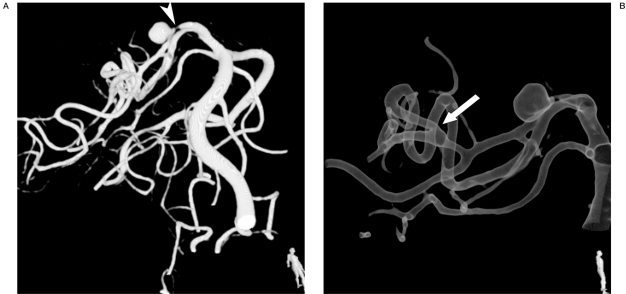
Figure 1.
Volume rendered (A) and gradient rendered (B) images obtained from 3DRA show dissecting aneurysm of P2 segment of the main trunk of right PCA. A P2 segment stenosis proximal to the aneurysm is present (A; arrowhead). A prominent posteromedial choroidal artery is identified with distal main PCA anastomosis at P3 level (B; arrow).

