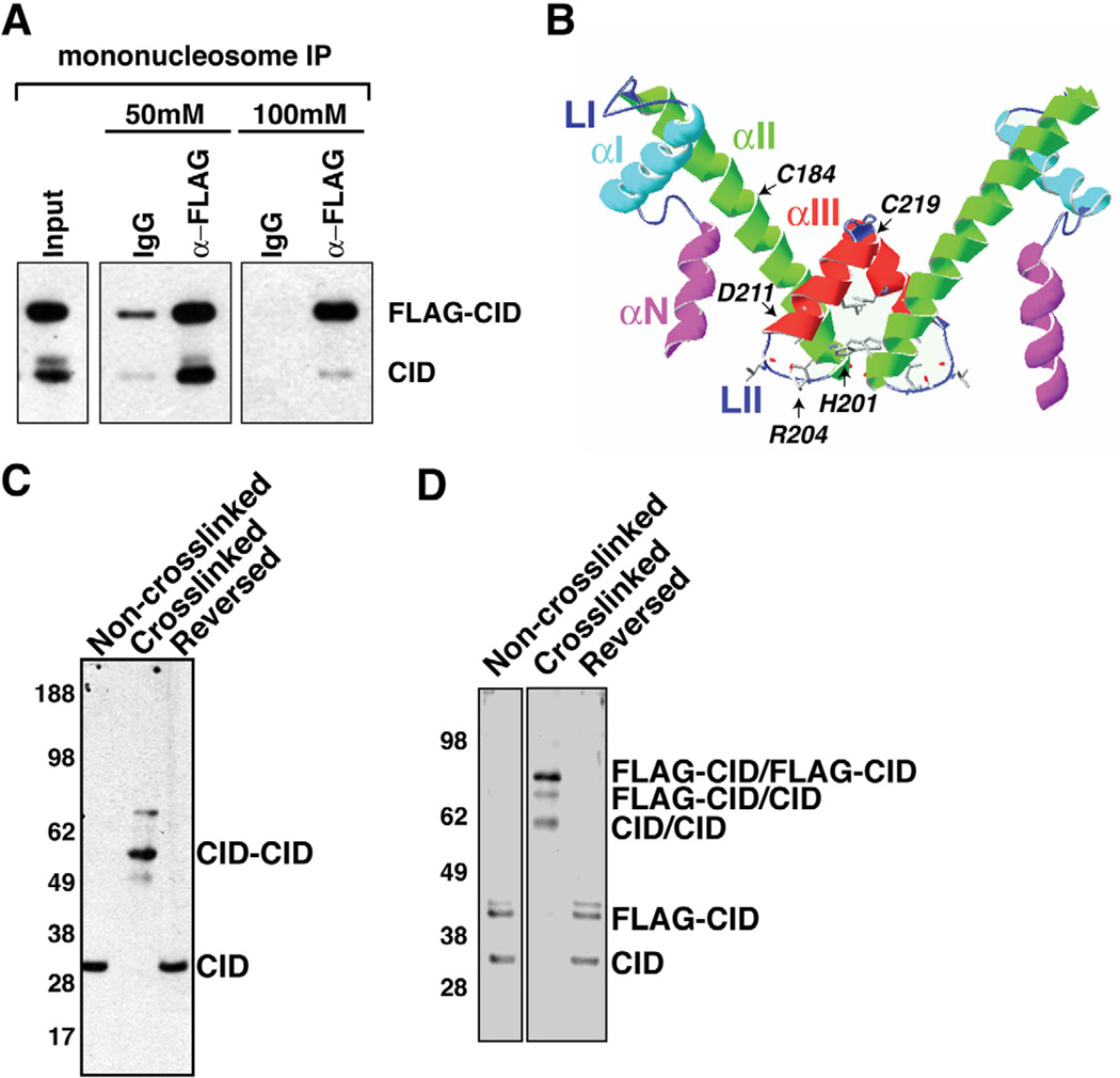
Figure 2. CID nucleosomes contain CID dimers.
- Endogenous CID coimmunoprecipitates with FLAG-CID from mononucleosomes. Inputs consist of mononucleosomes. α-mouse IgG beads were used in control IPs (IgG).
- Schematic display of the HFD of CID using a Cse4 model as reference (Bloom et al., 2006). Helices- αN, -αI, -αII, -αIII, and Loops LI, LII are indicated. CATD is comprised of LI and αII. The putative four helix bundle is comprised of the C terminal halves of αII and αIII.
- Cysteine crosslinking of S2 chromatin. CID monomers migrate at 30 kDa without crosslinking and after crosslink reversal. After crosslinking, the majority of CID migrate at 60 kDa, consistent with the size of dimers.
- Cysteine crosslinking of FLAG-CID chromatin. FLAG-CID and endogenous CID migrate as monomers before crosslinking and after crosslink reversal. After crosslinking, three major bands were detected by CID antibody that correspond to CID/CID, FLAG-CID/CID and FLAG-CID/FLAG-CID, respectively.
Also see Figure S2.

