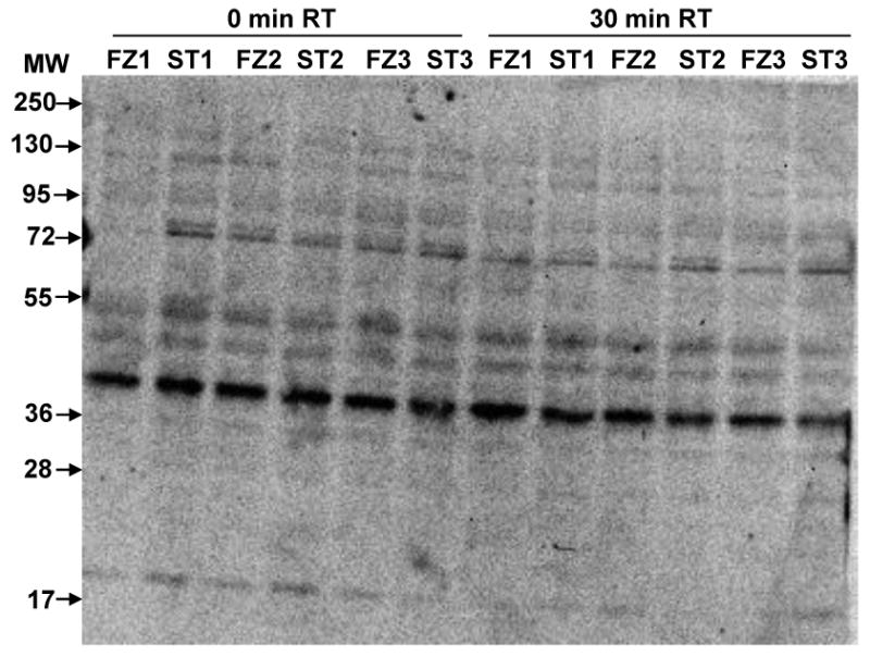
Figure 1. Patterns of phospho-serine appear similar in frozen and heat stabilized cortex.

Lysates were prepared after snap freezing in liquid nitrogen (FZ1-FZ3) or heat stabilization (ST1-ST3). Immediately prior to lysate preparation, brain regions were cut in half; one half was immediately placed in IEF buffer and processed (0 minutes room temperature (0 min RT)) and the other half was held for 30 minutes at room temperature (30 min RT) followed by buffer addition and processing. Gel membrane was screened with an antibody to phospho-serine.
