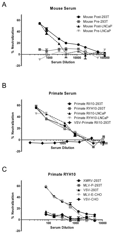
Fig. 1. Detection of XMRV Env neutralizing antibodies in positive controls.
MLV-luc(XMRV Env) pseudovirus infection of 293T/17 and prostate LNCaP cells was neutralized by sera from both mice (A) and rhesus macaques (B) challenged with XMRV, whereas no clear neutralization was observed with pre-immune sera, or HIV-luc(VSV G) pseudoviruses. (C) MLV-luc(MLV-E Env) pseudoviruses were neutralized by sera from rhesus macaques challenged with XMRV in mCAT-1 expressing CHO cells (CERD9 cells), but no clear neutralization of LacZ encoding MLV-P or HIV-luc(VSV G) pseudoviruses was observed in 293T/17 cells. Infection of pseudoviruses with firefly luciferase reporter was detected with Bright-Glo™ Luciferase Assay System (Promega), whereas infection of LacZ encoding MLV-P was measured using Galacto-Light Plus System for detection of β-Galactosidase (Applied Biosystems). Absolute values for the no sera controls were: MLV-luc(XMRV Env) gave 55810 RLU on 293T cells and 20213 RLU on LNCaP cells; HIV-luc(VSV-G) gave 65961 RLU on 293T cells and 51677 RLU on CHO cells; MLV-P gave 32356 RLU on 293T cells and MLV-luc(MLV-E Env) gave 41771 RLU on CHO cells. Results are presented as percentage of neutralization and shown as mean ± S.D. of triplicate measurements. A representative experiment of at least two experiments is shown.

