
Figure 2.
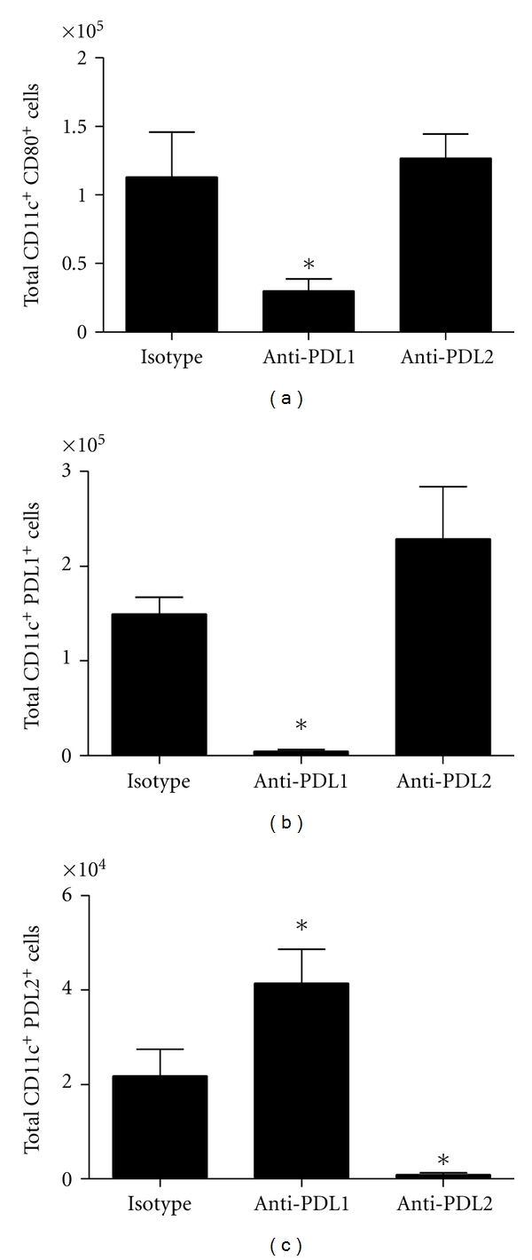
MLN CD11c+ leukocyte subpopulations following HSV-1 infection and treatment with anti-PD-L1 or anti-PD-L2 antibody. C57BL/6 mice were infected with 1,000 PFU HSV-1/cornea. At days 2, 4, and 6 p.i. mice were administered neutralizing antibody to PD-L1, PD-L2, or control IgG. At day 7 p.i. mice were exsanguinated and the MLN was processed for flow cytometry. (a) Total CD11c+ CD80+ cells. (b) Total CD11c+ PD-L1+ cells. (c) Total CD11c+ PD-L2+ cells. This figure summarizes three experiments (n = 9). Each bar represents the mean ± SEM. *P < 0.05 comparing the indicated group to the isotypic control antibody-treated group.