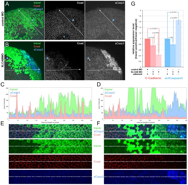
Figure 4. Cadherin junctional complex deficiency is the primary response to CRIM1 loss-of-function.
(A–B) Embryos were injected with either standard control MO or 15 ng each XLCAB MOs with dextran tracer (green), fixed at stage 13 and labeled for C-cadherin (Ccad) and activated caspase 3 (aCasp3). In (A) the asterisk indicates a region where labeling in the apical junctional complex was not imaged due to the optical sectioning plane. In (A and B), the blue arrowheads indicate isolated dying cells labeled positive for activated caspase 3. (C–D) Three-channel histograms indicating pixel intensity along a line interval of 450 pixels in control MO (A and C) and XLCAB MO (B and D) injected embryos. (E–F) Magnified images of regions along the line interval in (A and B) with color channel merge (top) and separated color channels corresponding to different labels as indicated. (G) Quantification of relative expression levels of C-cadherin and activated Caspase 3 in standard control MO and XLCAB injected embryos. The expression levels are determined by the ratio of average pixel intensities over 150-pixel intervals in tracer-positive regions and tracer-negative regions within the same embryo. In XLCAB injected embryos, relative expression levels were measured over intervals placed exclusively in tracer-positive, adherent regions or tracer positive, non-adherent regions (n = 8 for all categories).

