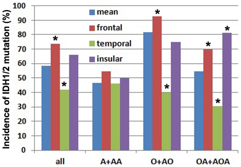
Figure 3. Gliomas of frontal and temporal origin had significantly different incidences of IDH1/2 mutation irrespective and in respective of tumor pathology.
The regional incidences of IDH1/2 mutation in all gliomas of the region irrespective of pathology were labeled with “All”. The regional incidences of IDH1/2 mutation in respective of pathology were labeled with “A+AA, O+AO and OA+AOA” respectively. *The incidence of IDH1/2 mutation in this region is significantly higher or lower than that in other regions (p<0.05, chi-square test).

