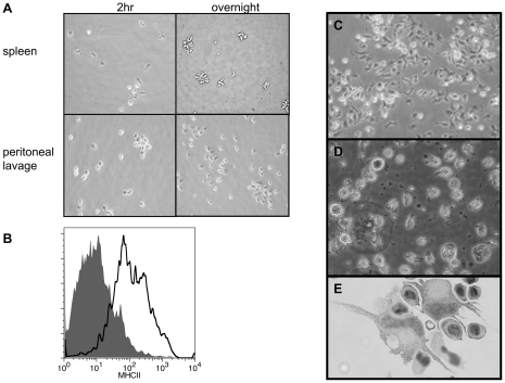
Figure 8. Isolation of tDC-like cells from the spleen and culture from peripheral blood mononuclear cells.
(A) Phase contrast images of tDC-like cells isolated from spleen using a mammalian protocol. Isolated cells are initially adherent (2 hr) but become non-adherent overnight, unlike macrophages from peritoneal lavage that remain adherent. (B) Non-adherent cells isolated from spleen express surface MHCII (grey filled histogram: pre-immune serum, black line histogram: anti-MHCII hyper-immune serum). (C) Phase contract image of adherent cells from mononuclear fraction of peripheral blood after 2 hr incubation. (D) Phase contrast of typical culture of adherent peripheral blood cells (20×, day 18). (E) Cytospin of peripheral blood cultures reveal the presence of large cells with long veils and kidney shaped nuclei (40×, day 7).

