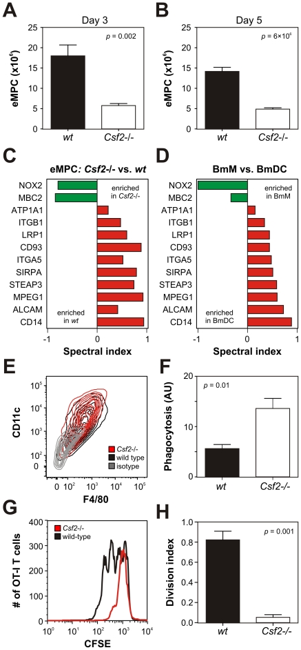
Figure 5. Analysis of eMPCs harvested from wild-type and GM-CSF-deficient (Csf2−/−) mice.
eMPCs isolated from wild-type (wt) and Csf2−/− mice were interrogated for cell number, function, and protein expression. Panel A–B: Accumulation of eMPCs 3 days (Panel A) and 5 days (Panel B) following intraperitoneal injection with thioglycolate. Results (N = 6) are means and SEMs. Panel C: Plasma membrane proteomic analysis of eMPCs isolated from Csf2−/− and wild-type mice. Differentially-expressed proteins were identified using the t-test and G-test (p<0.05 and G-statistic >1.5) and quantified using the spectral index. Panel D: Proteins differentially expressed by eMPCs isolated from Csf2−/− mice (see Panel C) were measured in BmMs and BmDCs and quantified using the spectral index. Panel E: Cell surface CD11c and F4/80 expression on eMPCs was assessed by flow cytometry. Results are presented as contour plots with 10% probability increments. Panel F: Phagocytosis of fluorescein-labeled E. coli by eMPCs. Results (arbitrary units, AU; N = 4) are means and SEMs. Panel G–H: Antigen cross-presentation by eMPCs. Ovalbumin (0.2 mg/mL)-treated eMPCs were incubated with CFSE-labeled spleen cells isolated from OT-I transgenic mice. Levels of CFSE were assessed in OT-I T cells selected by flow cytometry and expression levels of CD8 and Vb5 (Panel G). The division index was calculated using FlowJo software. Results (N = 4) are means and SEMs (Panel H). Where applicable, p-values were derived using a two-tailed Student's t-test. Results obtained for eMPC quantification, flow cytometry, phagocytosis and antigen cross-presentation are representative of 3 independent analyses.

