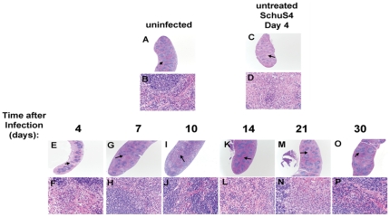
Figure 4. Pathological changes in the spleens of SchuS4 infected mice following antibiotic therapy.
Mice were infected intranasally with 50 CFU SchuS4. Three days after infection mice were treated once daily with 5 mg/kg LVF diluted in 5% dextrose water intraperitoneally for 14 days. At the indicated time points, spleens from untreated, SchuS4 infected animals (C and D) and SchuS4 infected mice treated with LVF (E–P) were fixed, sectioned, strained with H&E and assessed for pathological changes. Uninfected mice (A and B) served as normal controls. Representative photomicrographs for each time point are shown. Plates A, C, E, G, I, K, M, and O are at 2× magnification and plates B, D, F, H, J, L, N, P are 40× magnification of the areas indicated by the arrows on the 2× plates.

