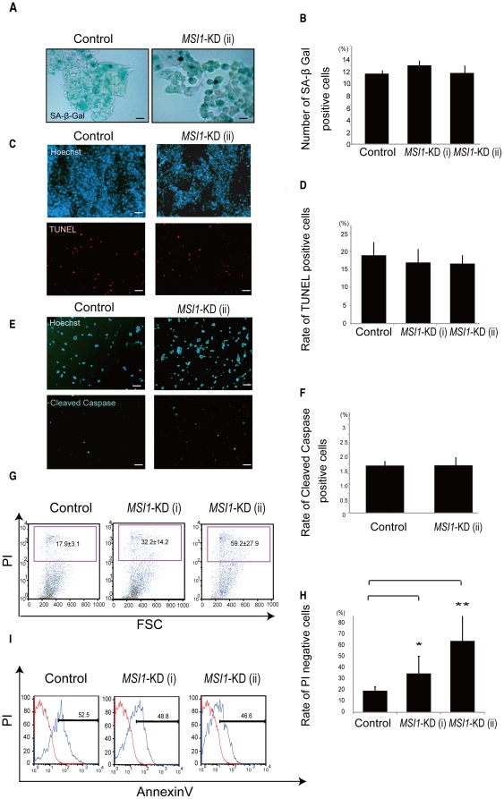
Figure 5. MSI1-KD cells may undergo non-apoptotic cell death.
(A and B) SA-β-Gal staining was performed to assess the senescence in controls and MSI1-KD cells. (A) Representative images of control cells and MSI1-KD cells stained for SA-β-Gal (Bar = 200 µm). (C, D, E, and F) Colonies were dissociated, and the cells were fixed to coverslips and stained for TUNEL in U251 MG cells (C and D), and for cleaved Caspase-3 in U251MG cells (E and F). Cell counts are plotted as bar graphs. An average of 25 high power fields was counted. (G and H) The number of PI-positive cells (indicating dead cells) was greater in the MSI1-KD cell population than in the control cell population. Error bars represent SEM. *P<0.01.**P<0.01. (I) The number of annexinV-positive in the MSI1-KD cells (indicating apoptotic cells) was not different from that in the control cell population.

