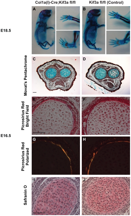
Figure 2. Kif3a expression in osteoblasts and osteocytes is not critical for embryonic skeletal development.
(A,B) Whole mount Alizarin Red (bone) and Alcian Blue (cartilage) staining of E18.5 Colα1(I) 2.3-Cre;Kif3afl/f l (A) and control (B) embryos. The size and limb patterning of Colα1(I) 2.3-Cre;Kif3afl/fl mice was similar to that of the control mice. (C,D) Movat's pentachrome staining of cross-sections of the radial/ulnar growth plates (cartilage-blue) in E16.5 Colα1(I) 2.3-Cre;Kif3afl/f l (C) and control (D) mice. (E–J) Cross-sections of E16.5 long bones stained with Picrosirius red (E,F-bright field; G,H-polarized light) to illuminate collagen and Safranin O (I,J) to demarcate cartilage. Both control and Colα1(I) 2.3-Cre;Kif3afl/f l mice have similar patterns of osteogenic and chondrogenic differentiation. Scale bar: 100 µm.

