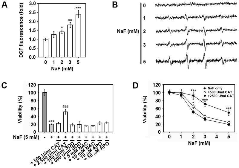
Fig. 4. Intracellular ROS is not directly related to NaF-induced reduction of mESC viability.
Cells were exposed to increasing concentrations of NaF (0–5 mM) for 24 h and then processed for flow cytometric analysis after (A) DCFH-DA staining and (B) ESR measurements. (C) Cells were incubated with 5 mM NaF in the presence and absence of various antioxidants for 24 h and then processed for the WST-8 assay. (D) Cells were incubated in the presence of NaF (0–5 mM) with and without 500 U/ml CAT or 2,500 U/ml CAT 24 h before determination of viability. *p < 0.05, **p < 0.01, and ***p < 0.001 vs. the untreated controls. ###p < 0.001 vs. 5 mM NaF treatment alone.

