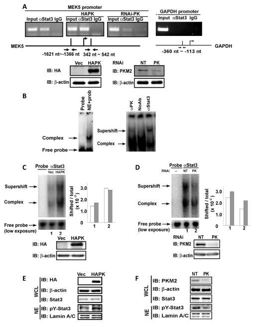
Figure 3. PKM2 upregulates MEK5 transcription by promoting stat3 DNA interaction and phosphorylation of stat3.
(A) (Upper panels) ChIP of the MEK5 promoter (MEK5 promoter) using antibody against stat3 in SW620 cells (αStat3). The cells were treated non-target RNAi (left, NT) or with RNAi target PKM2 (right, RNAi-PK), or HA-PKM2 was expressed in the cells (middle, HAPK). Inputs were PCR products from DNA extracts without ChIP. The primer pair positions are indicated. ChIP using rabbit IgG (IgG) was a negative control. ChIP targeting GAPDH promoter (GAPDH promoter) using antibody against stat3 was another negative control. (Lower panels) the cellular PKM2 (right) and HA-PKM2 (left) levels in SW620 cells that were treated with RNAi target PKM2 (PK) or with non-target RNAi (NT), or infected with virus that carry HA-PKM2 expression vector (HAPK) or vector alone (Vec) were analyzed by immunoblots using anti-HA antibody (IB:HA) or anti-PKM2 antibody (IB:PKM2). (B) DNA-protein complex (Complex) assembled on a 32P-labeled oligo containing the stat3 targeting sequence in nuclear extracts of SW620 cells was detected by gel-shift. Free probe indicates the 32P-labeled oligo probe without addition of nuclear extracts. The antibodies against PKM2 (αPK), stat3 (αStat3), or no antibody (NoAb) was added to the complex to create supershift (Supershift). (C) & (D) Supershift complex assembled with the 32P-labeled oligo and anti-stat3 antibody (αStat3) in the nuclear extracts of SW620 cells in which (C) HA-PKM2 was expressed (HAPK) or (D) PKM2 was knocked down (PK) was detected by gel-shift. Probe only (Probe) is the free probe without addition of nuclear extracts. The free probe (low exposure) is the loading control with 1/10 of exposure time in autoradiography. The quantification of the assembled complex and super shift complex were presented as percentage (Shifted/total ×102) of probe in the shift complex calculated by intensities of complex [complex (grey bars) or supershift (open bars)] divided by Intensities of total [free probe + complex + supershift). Immunoblots at bottom of each panel indicate levels of HA-PKM2 (IB:HA) and endogenous PKM2 (IB:PKM2) in the cells from which the extracts were prepared for the above gel shift experiments. (E) & (F) The levels of Y705 phosphorylated stat3 (IB:pY-Stat3) in the cell nucleus were analyzed by immunoblot of nuclear extracts (NE) of SW620 cells in which HA-PKM2 was expressed (E. HAPK) or the PKM2 was knocked down (F, PK). The total cellular stat3 levels were analyzed by immunoblot analyses of stat3 (IB:Stat3) in whole cell lysate (WCL). In (E), immunoblot of HA-tag (IB:HA) indicates the HA-PKM2 expression levels in the cells. In (F), immunoblot of PKM2 (IB:PKM2) represents cellular PKM2 levels in the cells. Immunoblot of lamin A/C in (E) and (F), and β-actin in (A), (C), (D), (E), and (F) are the loading controls. NTs in (A), (C), (D), (E), and (F) mean the cells were treated with non-target RNAi. Vec in (A), (C), (D), (E), and (F) means the cells were infected with virus that carry the empty vector.

