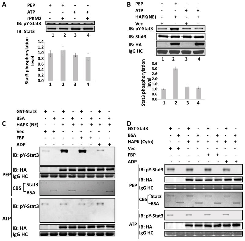
Figure 4. Phosphorylation of GST-stat3 by the rPKM2.
Phosphorylation of GST-stat3 by the rPKM2 (A) and HA-PKM2 (HAPK(NE)) immunopurified from nuclear extracts of SW620 (B) in the presence of 5 mM ATP (ATP) or 5 mM PEP (PEP) was revealed by immunoblot assays using antibody against Y705 phosphorylated stat3 (IB:pY-stat3). Immunoblot analyses using antibody against stat3 (IB:Stat3) indicates the amounts of GST-stat3 used in each reaction. The bottom panels in (A) & (B) are the quantitative analyses of immunoblot signals. The error bars represent the standard deviations of four measurements. Phosphorylation of GST-stat3 by the HA-PKM2 immunopurified from the nuclear (HAPK (NE) in C) and the cytoplasmic (HAPK(Cyto) in D) extracts of SW620 cells in the presence of 5 mM ATP (ATP) or 5 mM PEP (PEP) was revealed by immunoblots using antibody against Y705 phosphorylated stat3 (IB:pY-stat3). The reactions were also carried out in the presence/absence of 5 mM FBP (FBP), or 5 mM ADP (ADP). The immunoblot of HA (IB:HA) indicates the amounts of HA-PKM2 used in each reaction. IgG HC is the ponceau S stain of antibody heavy chain, representing the amounts of antibody used in immunopurification of HA-PKM2. Coomassie blue staining (CBS) indicates the amounts of GST-stat3 and BSA used in each phosphorylation reaction. Vec were the cells infected with virus that carry an empty vector.

