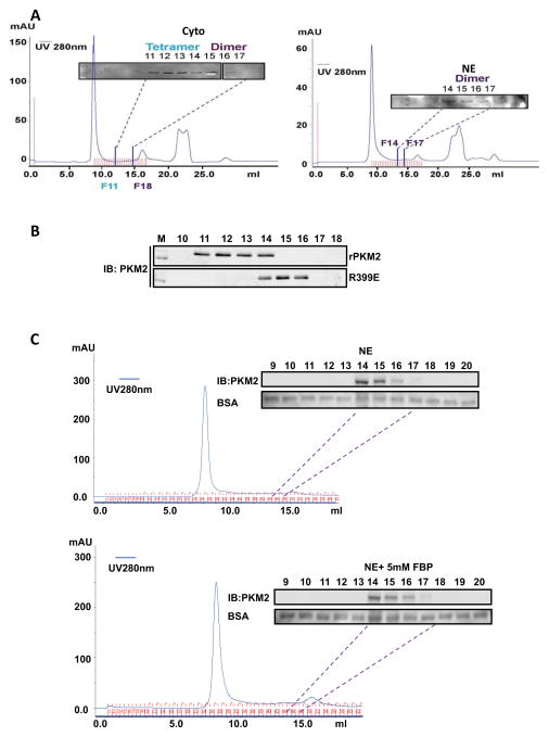
Figure 5. Dimer and tetramer PKM2.
(A) Chromatography fractionation of nuclear (NE, right) and cytoplasmic (Cyto, left) extracts of SW620 cells. The fractions were collected at 300 μl per fraction. The fractions 6 to 17 from cytoplasmic extracts and 9 to 18 from nuclear extracts were immunoblotted using the antibody PabPKM2. Fraction 11 (F11) co-eluted with 240 kDa and fraction 14 (F14) co-eluted with 120 kDa on the same column under identical conditions determined by chromatography molecular weight calibration standard (see Fig. S3 C&D). (B) Chromatography fractionation of the wild-type rPKM2 and R399E mutant. The fractions F10 – F18 were collected. The fractions were analyzed by immunoblot using the antibody PabPKM2 (IB:PKM2). (C) Effects of FBP on the dimer/tetramer status of PKM2 in nuclear extracts. Chromatography fractionation of nuclear (NE) extracts of SW620 cells treated (bottom panel) or untreated (upper panel) with FBP (5mM). The fractions were collected at 300 μl per fraction. The fractions 6 to 20 from nuclear extracts were immunoblotted using the antibody PabPKM2 (IB:PKM2). Immunoblots of BSA are the loading controls. 2 μg of BSA was added to each fraction. After SDS-PAGE separation of 20 μl of each fraction, the BSA was analyzed by immunoblot (BSA).

