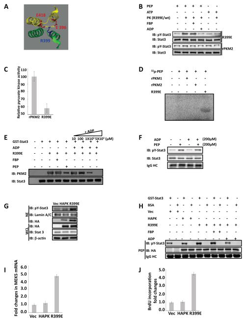
Figure 6. Dimeric PKM2 is active protein kinase and expression of the R399E mutant promotes cell proliferation.
(A) Part of x-ray crystal structure of human PKM2. The structure was obtained from PDB bank DOI: 10.1021/bi0474923. The residue R399 and its interactive residues E418, D357, and E396 are highlighted in color. (B) Phosphorylation of GST-stat3 by 10 μg/ml of rPKM2 (rPKM2) and R399E mutant (R399E) in the presence of 5 mM ATP (ATP), 5 mM PEP (PEP), 5 mM FBP (FBP), and/or 5 mM ADP (ADP) was revealed by immunoblot assays using antibody against Y705 phosphorylated stat3 (IB:pY-stat3). Immunoblot analyses using antibody against stat3 (IB:Stat3) indicates the amounts of GST-stat3 used in each reaction. (C) Pyruvate kinase activity of the rPKM2 or R399E (5 μg/ml) was analyzed by the method described by Christofk and coworkers. The pyruvate kinase activity was expressed as relative pyruvate kinase activity by define the activity in the rPKM2 as 100. (D) Phosphorylation of GST-stat3 by 10 μg/ml of rPKM2, the rPKM1, or the R399E in the presence of 32P-PEP (~0.002 μCi) and unlabeled PEP (5 mM). The reaction mixture were separated by SDS-PAGE and subjected to autoradiograph. (E) Interaction of GST-stat3 and R399E in the presence of FBP (5 mM), PEP (5 mM), or various concentrations of ADP (inducated) was analyzed by GST-pull-down. The co-precipitation of R399E with GST-stat3 was detected by immunoblot using the antibody against PKM2 (IB:PKM2). Immunoblots of precipitates using antibody against stat3 (IB:Stat3) indicate the amounts of GST-stat3 that was pulled-down by the glutathione beads. (F) Phosphorylation of GST-stat3 by the PKM2 (10 μg/ml) purified from nuclear extracts of SW620 cells in the presence of 200 μM PEP (PEP) and 200 μM ADP (ADP) was revealed by immunoblot assays using antibody against Y705 phosphorylated stat3 (IB:pY-stat3). Immunoblot analyses using antibody against stat3 (IB:Stat3) indicates the amounts of GST-stat3 used in each reaction. (G) Phosphorylation of stat3 in SW480 cells was examined by immunoblot analyses of the nuclear extracts (NE) using antibody against the Y705 phosphorylated stat3 (IB:pY-Stat3). PKM2 (HAPK) or the R399E (R399E) was expressed in the cells. Immunoblots using anti-HA antibody (IB:HA) and anti-stat3 antibody (IB:Stat3) in the whole cell lysate (WCL) indicate the levels of stat3 and HA-PKM2/HA-R399E in the cells. Immunoblot of lamin A/C (IB:Lamin A/C) and β-actin (IB:β-actin) are loading controls. (H) Phosphorylation of GST-stat3 by the HA-p68 and the HA-R399E that were immunopurified from cell lysate of SW480 cells in the presence of 5 mM PEP was revealed by immunoblot analyses using antibody against the Y705 phosphorylated stat3 (IB:pY-Stat3). The reactions were also carried out in the presence/absence of 5 mM FBP (FBP), or mM of ADP (ADP). The immunoblot of HA (IB:HA) indicates the amounts of HA-PKM2 or HA-R399E used in each reaction. (I) Expression of MEK5 mRNA in SW480 cells was analyzed by RT-PCR. HA-PKM2 (HAPK) or HA-R399E (R399E) was expressed in the cells. The results are presented as fold changes in PCR products before and 48 hours after HA-PKM2 or HA-R399E expression. (J) Cell proliferations of SW480 cells were measured using a proliferation kit. Proliferations were presented as fold changes in BrdU incorporation before and 48 hours after HA-PKM2 (HAPK) or HA-R399E (R399E) expression. The BrdU incorporation of the cells that were transfected with the empty vector (Vec) was defined as 1. In (F) and (H), IgG HC is the ponceau S stain of antibody heavy chain, representing the amounts of antibody used in immunopurification of HA-PKM2 from the extracts. Error bars in (C), (I), and (J) are standard deviations of three independent measurements. Vec in (G), (H), (I), and (J) are the cells transfected with the empty vector.

