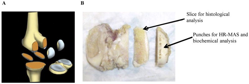
FIG 1.
Cartilage specimen collection from advanced OA patients during TKA surgery (A). The diagram of the five specimens containing bone and cartilage sectioned during surgery: lateral/medial inferior femoral condyles (LIFC/MIFC), lateral/medial posterior femoral condyle (LPFC, MPFC) and tibial plateau containing lateral/medial tibia (LT/MT). (B) Location of the punches taken for HR-MAS and the slice for histology. Picture taken from [29].

