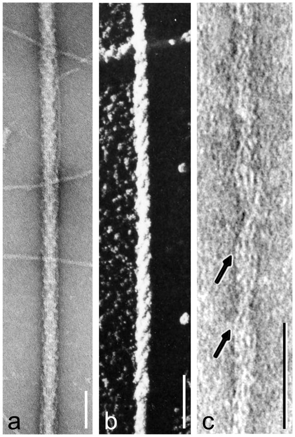
Figure 2.
Negative staining and shadowing. (a) unpurified tarantula thick filaments, with thin filaments in background (from [4]); (b) metal-shadowed tarantula thick filament showing right-handed helical tracks (from [5] with permission); (c) negatively stained thin filament from frog cardiac muscle (from [103] with permission); this vertebrate image is chosen for the clarity with which it reveals tropomyosin (arrowed strands), although it was invertebrate filaments that first demonstrated the tropomyosin-shift mechanism unequivocally [12]. In (a,c), protein is white and stain dark; in (b) metal is white. Scale bars: a, b (100 nm), c (50 nm).

