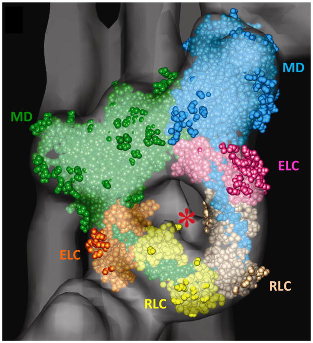
Figure 6.
Fitting of myosin head atomic structure into repeating J-motif in tarantula reconstruction (Fig. 5e). Motor domain (MD), essential light chain (ELC) and regulatory light chain (RLC) of blocked head are in green, orange and yellow; those of free head are blue, pink and beige. Asterisk indicates rod-like volume of density that represents the start of the S2 portion of the myosin tail. Modified from [50].

