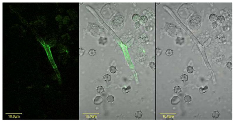
Fig. 9.

A C. albicans hypha from murine kidney tissue immunolabeled with anti-Als4. A BALB/cByJ mouse was inoculated via the lateral tail vein with 5 × 10⁵ cells of C. albicans strain CAI12. At 28 h post-inoculation, the kidneys were removed, minced with a razor blade, and homogenized. A portion of the supernatant was treated with anti-Als4 and a FITC-conjugated secondary antibody. Cells were imaged using an Olympus BX50 FluoView microscope. Images above are illuminated with laser only (488 nm; left panel), white light (right panel) or both (center panel). Als4-positive cells were difficult to find, suggesting that they were rare in this model. Repetition of the experiment with other mice yielded the same result.
