
Fig. 1.
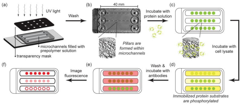
Schematic of hydrogel post fabrication, protein immobilization, kinase assay, and detection of substrate phosphorylation. (a) Hydrogel pillars are photopatterned from PEG prepolymer solutions containing acryloyl-functionalized NHS within microchannels. (b) Photograph of channels with pillars. Unreacted prepolymer solution is washed from the channel leaving polymerized pillars within the microchannels. (c) Protein substrate solution is flowed into the channels and allowed to react with NHS groups in the hydrogel pillars. Unincorporated protein is washed from the channels and (d) a kinase reaction mixture containing cell lysate is flowed into the channels. (e) Phosphorylated substrate is labeled with fluorophore-conjugated antibodies and (f) imaged with epifluorescence microscopy.