
Fig. 3.
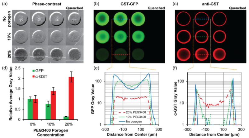
Immobilization of GST-GFP in 5% PEG700DA hydrogels with 0–20% PEG3400 porogen. (a) Phase-contrast images of hydrogel pillars. Pillars were incubated with 2 μg/μL GST-GFP and subsequently incubated with anti-GST and Alexa Fluor 594-conjugated secondary antibody. Fluorescence images showing (b) GST-GFP and (c) anti-GST with Alexa Fluor 594-conjugated secondary antibody localization throughout the pillars. The first three columns are triplicates and pillars shown in the last column were quenched with 0.5 M hydroxylamine prior to exposure to GST-GFP. Contrast is enhanced in image to emphasize low-signal areas. (d) Fluorescence intensity of GST-GFP and anti-GST relative to those detected in hydrogels lacking porogen. Error bars = SD (n ≥ 4 pillars). (e) Typical GST-GFP and (f) anti-GST fluorescence intensity profiles along a diameter of the hydrogels pillars. Gray value range = 0–255.