
Fig. 4.
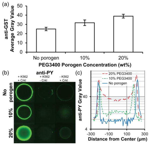
Immobilization of GST-Crkl and detection of K562 cell lysate-mediated phosphorylation in 5% PEG700DA hydrogels with 0–20% PEG3400 porogen. (a) Average gray values for anti-GST detection of GST-Crkl after incubating pillars in a solution of 4 μg/μL GST-Crkl and exposure to K562 cell lysate. Error bars = SE (n ≥ 3 experiments). (b) Images of anti-phosphotyrosine signal (overexposed to emphasize low signal areas) in these pillars. Pillars shown in the middle and last column were negative controls and any observable signal suggests nonspecific cell lysate adsorption (middle column, no GST-Crkl immobilized) or nonspecific antibody adsorption (last column, no K562 cell lysate). (c) Typical fluorescence intensity profiles of anti-phosphotyrosine along the diameters of the representative pillars shown in (b), first column. Gray value range = 0–255.