
Figure 6.
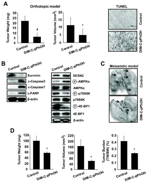
DIM-C-pPhOH inhibits tumor growth and lung metastasis in vivo. (A and B) The orthotopic mouse model of lung cancer. A549 cells were orthotopically implanted into athymic nude mice, and each mouse was dosed 3 times a week by oral gavage with either corn oil (control) or DIM-C-pPhOH (30 mg/kg/day) for 4 weeks starting 7 days after implantation. Median tumor weights and volumes (A, left panel) were calculated as described in the Materials and Methods. The data are presented as means with SD (n=10 per group). *P<0.05 and #P<0.001 vs control group. (A, right panel) TUNEL staining. Tumor sections were stained using the DeadEnd colorimetric kit as described in Materials and Methods. The apoptotic tumor cells are stained. Images were collected at high (×100) magnification. (B) Protein expression in tumor lysates. Tumor lysates from tumor samples were further analyzed by western blot analysis, and β-actin was used as a loading control. (C and D) The metastatic mouse model of lung cancer. A549 (2 × 10⁶) cells were inoculated into athymic nude mice via the tail vein for 4 weeks before treatment, and each mouse was dosed 3 times a week by oral gavage with either corn oil (control) or DIM-C-pPhOH (30 mg/kg/day) for 4 weeks. (C) Lung micrographs show development of multiple tumor foci (arrows). (D) Metastatic tumor weight, volume, and burden were calculated.