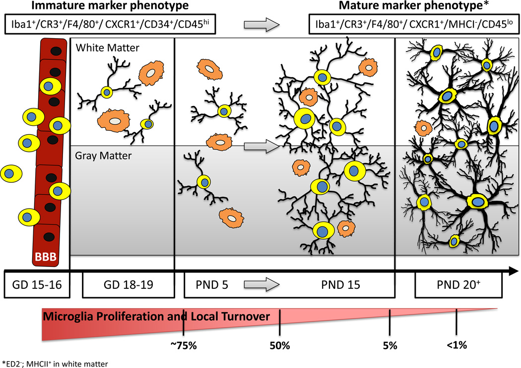
Figure 1.
Developmental Stages Leading to Establishment of a Mature Population of Brain Microglia. Prior to late gestation (GD15-16), bone marrow derived mononuclear cells cross the blood-brain barrier (BBB) and take up residence in the brain parenchyma. These cells typically possess little cytoplasm and have small or nonexistent appendages (indicated as process-free yellow cells with blue nuclei). At early stages of colonization (GD18–19), these cells are identifiable in white matter regions (or along vascular/ ventricular margins) possessing either an amoeboid/ phagocytic morphology (orange cells) or long, highly-branched processes. During early postnatal stages (~PND5), these highly proliferative mononuclear cells are observed in both white matter and gray matter regions of the brain with either morphological phenotype. During a critical period of postnatal microglia development (PND5– PND15), the number of microglia increases dramatically. During this time, an increased ratio of ramified versus amoeboid microglia becomes apparent, with the cells having noticeably more complex process arbors and cytoplasmic material. The microglia present towards the latter part of this maturation window (~PND15) are well distributed throughout the brain, facilitating surveillance of the majority of the parenchyma; they exhibit a significantly reduced proliferative capacity; they begin to exhibit more heterogeneity (both within and across brain regions) in terms of their morphology, orientation, and process field organization; and they begin to express a somewhat modified constellation of cell surface markers. These maturation features continue such that by PND20 the adult population of microglia is fairly well established. In the absence of stimulation, these cells are highly ramified with complex process arbors encompassing the entire brain parenchyma, and they possess little to no evidence of proliferation or turnover from the systemic population. * These marker lists are not comprehensive (e.g., mature microglia express ED2− and white matter microglia are MHCII+).

