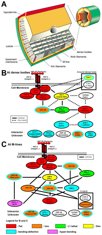
Figure 1.
Muscle structure and sets of interacting proteins found at muscle focal adhesions (M-lines and dense bodies) in C. elegans striated muscle. (A) Organization of the myofilament lattice in body wall muscle of C. elegans. The upper right depicts a portion of a nematode cut in cross section with body wall muscle quadrants in yellow. The enlargement shows the organization of filaments and membrane attachment structures in several planes of section. The sarcomeric region is confined to a narrow zone adjacent to the cell membrane along the outer side of the muscle cells. Note that all the dense bodies (Z-disk analogs) and M-lines are anchored to the muscle cell membrane and thus positioned to transmit the force of muscle contraction. (B and C) Sets of interacting proteins identified at dense bodies and M-lines that begin with UNC-52 (perlecan) in the ECM and integrins in the muscle cell membrane, and continue until the thin and thick filaments, respectively. Vertebrate homologs or orthologs are indicated in parentheses. The references for the dense body interactions are in reference [15], and unpublished data from the Benian and Moerman labs; the references for the M-line interactions are in references [4] and [15], and unpublished from the Benian lab. As shown at the bottom of the figure, the diagrams are color-coded to reflect loss of function mutant phenotype: red, Pat embryonic lethal; orange, Unc adults; yellow, Dim (disorganized myofibrils); green, L1 larval lethal; yellow, Dim (disorganized muscle); blue, bending-defective; purple, hyper-bending. The bending-defective (blue) and hyper-bending (purple) phenotypes were identified using the new locomotion assay described here. Note that mutants for five proteins, UNC-95, PKN-1, UNC-89, UNC-96, and UNC-82, are both Unc (orange) and bending defective (blue outer oval). DIM-1 is both Dim (yellow) and bending defective (blue outer oval). In addition to those proteins with Pat or L1 lethal phenotypes, bending assays were not conducted for TTN-1, CeTalin, paramyosin or CSN-5. HSP-25 was tested, but did not show a bending defect.

