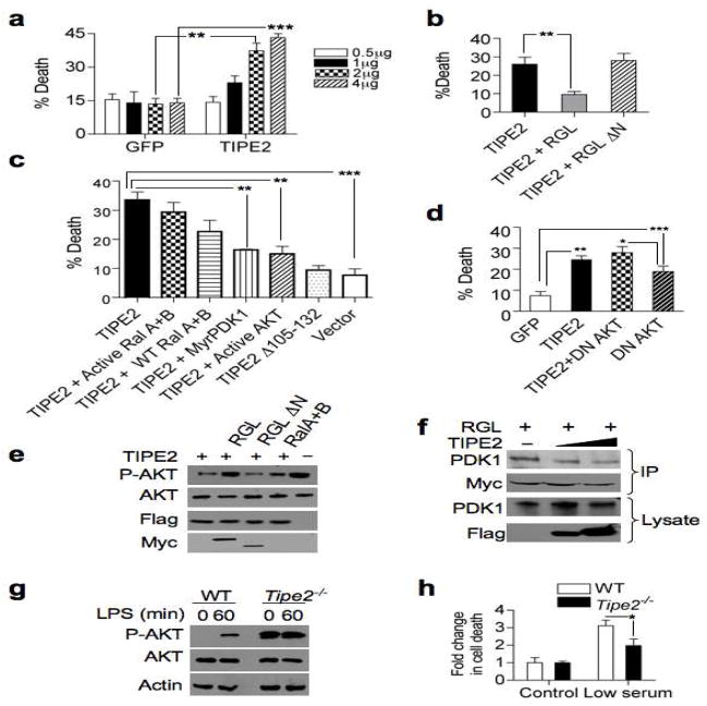
Figure 2. TIPE2 promotes cell death by inhibiting RGL-induced AKT activation.
(a) TIPE2 promotes cell death in 293T cells. Cells were transfected with the indicated amounts of TIPE2-Flag construct (TIPE2) or control GFP vector (GFP). After 24 hrs, cell death was assessed by trypan blue staining. Data shown are means ± S.E.M (n=3), and are representative of 5 independent experiments. ** p < 0.01; *** p < 0.001. (b) RGL rescues TIPE2-induced cell death. 293T cells were transfected with TIPE2-Flag together with RGL or RGL ΔN plasmids for 24 hrs, and cell death was assessed as in (a). Data shown are means ± S.E.M (n=5), and are representative of 5 independent experiments. ** p < 0.02. (c) TIPE2-induced death is mediated by the PDK1-AKT axis. 293T cells were transfected with TIPE2-Flag or TIPE2 Δ3105-132 plasmids, with or without activated RalA and RalB, wild type RalA and RalB, activated AKT, or myristoylated PDK1 plasmids for 24 hrs. Cell death was assessed as in (a). Data shown are means ± S.E.M (n=3). *** p< 0.001, ** p< 0.005. (d) AKT inhibition is responsible for TIPE2-induced cell death. 293T cells were transiently transfected with either GFP, TIPE2, dominant negative (DN) AKT, GFP plus TIPE2, or DN AKT plus TIPE2 plasmids. Cell death was assessed as in (a). Data shown are means ± S.E.M (n=5) of the cell death rates, and are pooled from 2 independent experiments. *** p < 0.001, ** p < 0.01, * p < 0.05. (e) TIPE2 reduces phospho (P)-AKT (S473) levels. 293T cells were transfected with the indicated constructs for 24 hours, and protein levels were determined by IB. (f) TIPE2 decreases RGL interaction with PDK1. Lysates of 293T cells transiently transfected with RGL-Myc (2 μg/10-cm plate) and increasing amounts of TIPE2-Flag constructs (5 μg to 10 μg/10-cm plate) were immunoprecipitated with anti-Myc, and subjected to SDS-PAGE and IB. (g) Increased phosphorylation of AKT in Tipe2−/− splenocytes. Wild type and Tipe2−/− splenocytes were treated with LPS (200 ng/ml) for the indicated times. Cell lysates were subjected to SDS-PAGE and IB. Data shown are representative of three independent experiments. (h) Tipe2−/− splenocytes are resistant to serum deprivation-induced death. Tipe2−/− or wild type splenocytes were incubated in DMEM containing 10% (control) or 0.2% (low serum) FBS for 4 hrs, and cell death was assessed as in (a). Data shown are means ± S.E.M (n=3), and are representative of three independent experiments. * p < 0.05.

