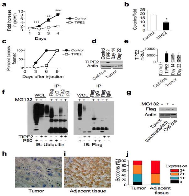
Figure 4. TIPE2 over-expression suppresses Ras-induced transformation whereas TIPE2 down-regulation is associated with human hepatocellular carcinogenesis.
(a) TIPE2 inhibits the growth of Ras-transformed cells. Ras-transformed NIH3T3 cells that did or did not express TIPE2 were cultured in DMEM containing 0.5% FBS. Cells were fixed at 24 hr intervals and stained with methylene blue. Results are means ± S.E.M (n=5) of relative absorbance with the value at day one set as 1, and are representative of three independent experiments. ** p<0.001; *** p < 0.0001. (b) TIPE2 inhibits soft-agar colony formation of Ras-transformed cells. Ras-transformed NIH3T3 cells that did or did not express TIPE2 were cultured in duplicates in soft-agar plates as described in Methods, and colonies were counted in 20 random fields for each culture under 40X magnification. Results are means ± S.E.M (n=20) and are representative of three independent experiments. * p < 0.0001. (c) TIPE2 delays tumorigenesis in nude mice. Ras-transformed NIH3T3 cells that did or did not express TIPE2 were injected subcutaneously into the rear flanks of nude mice (2×10⁶ cells/injection, n=3), and tumor formation was monitored daily. All sites injected with Ras-transformed NIH3T3 cells eventually developed tumors, whereas control NIH3T3 or NIH3T3-TIPE2 cell lines did not give rise to tumors during the course of this study. Data shown are percent of tumors formed, and are representative of two independent experiments. The differences between the two groups are statistically significant (p = 0.014). (d–e) Loss of TIPE2 protein expression in Ras-transformed NIH3T3 tumors. Tumors from two separate experiments were excised at day 14 or day 22. Expression of TIPE2-Flag protein (d) and mRNA (e) was examined by SDS-PAGE and IB, and qRT-PCR, respectively. Cell lines used in these assays were Ras-transformed NIH3T3 cells that did (TIPE2) or did not (control) express TIPE2-Flag. qRT-PCR data are shown as means ± S.E.M (n=3) with the value of the control group set as 1. (f) Ubiquitination of TIPE2. 293T cells were transfected with either TIPE2-Flag, or p50-Flag as a positive control. The cells were treated with MG132 (10μM), the lysates were immunoprecipitated with an antibody against Flag, and the ubiquitination status was examined as we previously described (Carmody et al., 2007). (g) TIPE2 protein is degraded in Ras-transformed NIH3T3 tumors. Reconstituted cells from Ras3T3-TIPE2 tumors were treated with MG132 for 24 hrs and TIPE2 protein levels were examined by Western blot and compared to untreated pre-injected cells. (h–j) Loss of, or decreased, TIPE2 expression in human hepatocellular carcinoma. TIPE2 expression in tumor tissue (h) and control hepatic tissue adjacent to the tumor (i) from the same patient was determined by immunohistochemistry as described in Methods. TIPE2-positive cells are shown in brown. Original magnification, x400. Quantitation of the TIPE2 signal for 116 patients was performed as described in Methods (j). The differences between the two groups are statistically significant (p < 0.001).

