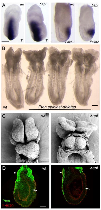
Figure 4. Conditional deletion of Pten in the epiblast shows that Pten acts in extraembryonic cells to control axis specification.
(A) The primitive streak marker T was expressed correctly in a single primitive streak in all Pten-epiblast deleted (Δepi) embryos examined (13/13), as was the anterior streak marker FoxA2. Scale bars = 250 μm. (B) The morphology of e8.5 Pten-epiblast deleted embryos, viewed from the dorsal side. All embryos of this genotype form a single anterior-posterior body axis (n>50). Like the wild-type embryo on the left, the three mutants (on the right) form somites and close the neural tube in the trunk, but have headfolds that fold in irregular patterns. Scale bar =250 μm. (C) SEM views of the ventral side of wild-type and Pten-epiblast deleted e8.5 embryos. In contrast to the single, looping heart tube in wild type, the mutant heart is composed of two tubes that have not fused on the midline (cardia bifida). Scale bar = 100 μm. (D) Pten protein localization in e6.5 wild-type and Pten-epiblast deleted embryos. Arrows indicate the epiblast; arrowheads indicate extraembryonic ectoderm. Pten is detectable in the epiblast, visceral endoderm and extraembryonic ectoderm of wild-type embryos (8/8). Pten protein is not detectable in the epiblast of Pten-epiblast deleted embryos (3/3), although it is still detectable in the extraembryonic ectoderm and visceral endoderm. Scale bar = 50μm.

