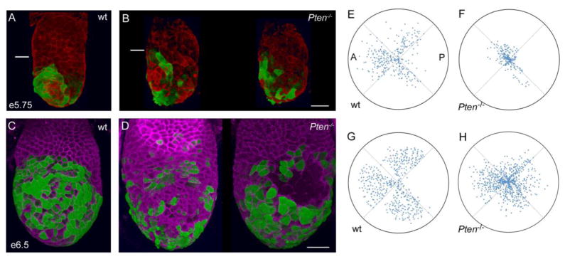
Figure 6. Pten mutant Hex+ AVE cells are dispersed around the embryo.
(A–B) 3D reconstructions of e5.75 embryos during AVE migration, showing expression of Hex-GFP (green, staining with anti-GFP antibody) and F-actin (phalloidin, red). White line marks the embryonic/extraembryonic boundary. (A) Wild type, a left/anterior view. Migration is in progress and Hex+ cells have not yet reached the boundary of the embryonic region. (B) The left and right sides a Pten−/− embryo. AVE cells are more dispersed in the mutant embryo. The leading cells have arrived at the embryonic/extraembryonic border, but many cells remain distally located. Scale bar for A and B = 40μm. (C, D) 3D reconstructions of e6.5 embryos after AVE migration, showing expression of Hex-GFP (green, staining with anti-GFP antibody) and E-cadherin (magenta). (C) Anterior view of wild-type; there are no AVE cells in the back (not shown) or at the distal tip of the embryo. (D) Two views of a Pten−/−embryo; posterior view to the left and anterior view on the right. AVE cells are present all sides of the embryo, and numerous cells remain distally located. (C) Scale bar for C and D = 50μm. (E–H) Polar plots representing the distribution of AVE cells in embryos like those in (A–D). Each dot represents the position of one Hex+ cell. Plots are oriented so the most populated quadrant (the presumptive anterior) is oriented to the left. The position of each cell is indicated both with respect to its position around the circumference of the embryo (angle) and distance migrated along the proximal-distal axis (proximity to center of the plot). Data from 4 wild-type embryos (+/+) at e5.75 (E) and 4 wild-type embryos (+/+) at e6.5 (G) reveal that most Hex+ cells are in the 3 anterior quadrants and have moved away from the distal tip. Data from 3 Pten−/− mutant embryos at e5.75 (F) and 5 Pten−/− mutant embryos at e6.5 (H) indicate that cells are more evenly distributed among the quadrants and many more remain near the distal tip. Heterozygous embryos were excluded from this analysis, as they appear to have an intermediate behavior between +/+ and Pten−/− embryos (Supp. Table S2).

