
Figure 2.
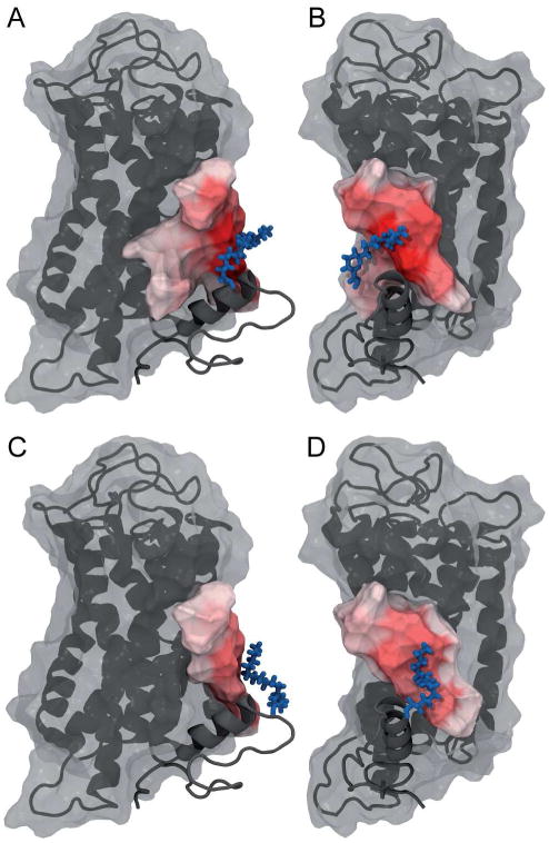
Depiction of the amino acids which experience close contacts with the lipid modification attached to Cys322 (panels A and B) and to Cys323 (panels C and D) shown from two different angles. The membrane extracellular side is up and the cytoplasmic side is down. Rhodopsin is shown with a semitransparent surface. All amino acids that experience contacts are shown in a nontransparent surface representation. The intensity of the red color indicates the number of contacts with the lipid modification. The palmitoylation at Cys322 shows many contacts with helix H1 and some with helix H7. The palmitoylation at Cys323 shows a similar pattern, but with a narrower distribution and fewer contacts overall. The figure was prepared using VMD67.