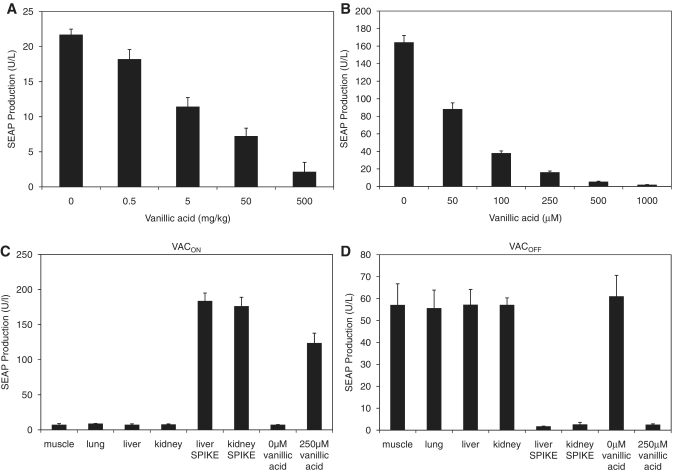
Figure 5.
Vanillic acid-controlled SEAP expression in mice. (A) CHO-VAC12 cells were microencapsulated in alginate-poly-(l-lysine)-alginate beads and implanted intraperitoneally into female OF1 mice (4 × 10⁶ cells per mouse). The implanted mice received different concentrations of vanillic acid twice daily. Seventy-two hours after implantation, the level of SEAP in the serum of the mice was determined. Data represent mean ± SEM of 8 mice per treatment group. (B) SEAP expression profiles of the microencapsulated CHO-VAC12 implant batch were cultivated in vitro for 72 h at different vanillic acid concentrations. (C and D) Extracts of wild-type mouse organs were assessed for their vanillic acid content based on their ability to induce the (C) VACOFF or (D) VACON systems. Vanillic acid-spiked organs were used as positive control. All samples were compared to the effect of 250 μM vanillic acid to show the fully induced state of the systems. All extracts were added to CHO-K1 cells transiently transfected with either the VACON or the VACOFF systems and SEAP expression was assessed after a cultivation period of 48 h.

