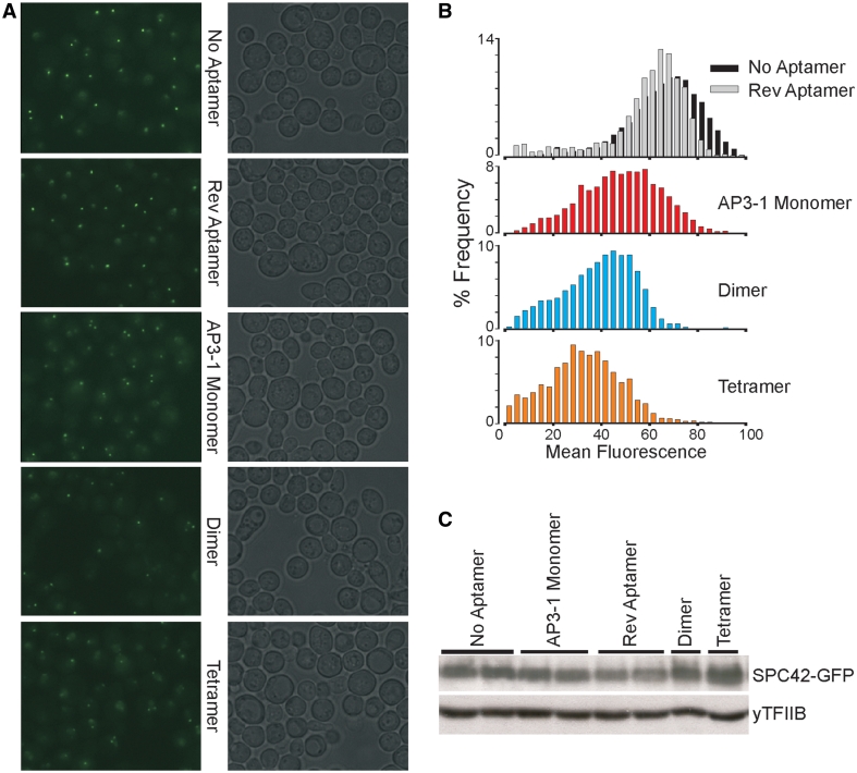
Figure 8.
AP3 inhibits GFP fluorescence in vivo. (A) Fluorescence microscopy of yeast cells that co-express SPC42-GFP and the optimized short AP3. Each fluorescence image is paired with a differential interference contrast image. Monomeric, dimeric and tetrameric AP3 expressions were tested, with monomeric AP3-1 in reverse orientation or no aptamer as negative controls. Expression of optimized short AP3 decreased the local fluorescence intensity and dimer spindle pole bodies were observed as the copy number of aptamer coding sequence increase. (B) Histogram of the fluorescent signals from experiments shown in (A). Mean fluorescence intensity decreased as the copy number of AP3 coding sequencing increase. (C) Confirming SPC42-GFP expression with western blot analysis. Altered expression of GFP in different strains does not explain the results of (A) and (B).

