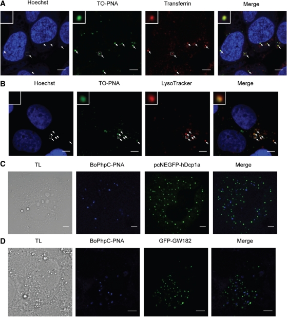
Figure 6.
(A) Confocal microscopy of Huh7 cells co-incubated with 3 µM Cys-K-(TO)PNA-K3 and 50 µg/mL Alexa 594-conjugated human transferrin for 1.5 h. (B) Confocal microscopy of Huh7 cells treated with 3 µM Cys-K-(TO)PNA-K3 observed at 24 h after PNA treatment; lysosomes were stained using LysoTracker Red. (C) Representative image of confocal microscopy of Huh7 cells expressing P-body marker (pcNEGFP-hDcp1a) treated with 3 µM BoPhpC-PNA. (D) Representative image of confocal microscopy of Huh7 cells expressing GW-body marker (GFP-GW182) treated with 3 µM BoPhpC-PNA.

