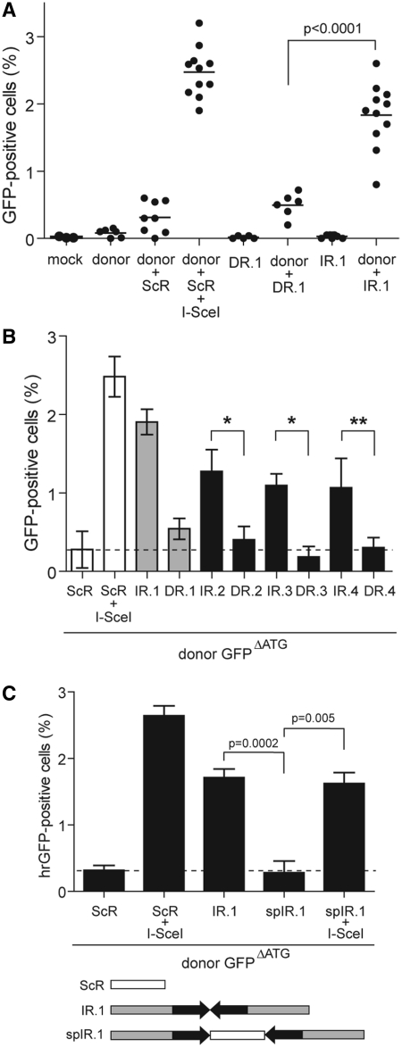
Figure 3.
Effect of repetitive DNA sequences in a direct or inverted repeat configuration on homology-directed gene repair in HeLa cells. (A) HeLa cells were transfected with acceptorDR.1 or acceptorIR.1 alone (DR.1 and IR.1, respectively) or with either of these two acceptor plasmids in combination with the donor construct GFPΔATG (donor + DR.1 and donor + IR.1, respectively). Mock-transfected HeLa cells (mock) and HeLa cells transfected with GFPΔATG alone (donor) or together with acceptorScR (donor + ScR) served as negative controls. The positive control for the rescue of GFP expression by HR was provided by cotransfecting HeLa cells with acceptorScR, GFPΔATG and the I-SceI-encoding plasmid pCAG.I-SceI (donor + ScR + I-SceI). Quantification of the number of GFP-positive cells was carried out by flow cytometry at 4 days post-transfection. A minimum of 5 and a maximum of 11 independent experiments were performed with 10 000 events corresponding to viable cells being measured per sample. (B) HeLa cells were cotransfected with GFPΔATG plus either acceptorIR.2, acceptorDR.2, acceptorIR.3, acceptorDR.3, acceptorIR.4 or acceptorDR.4. To facilitate comparison, data sets corresponding to HeLa cells cotransfected with GFPΔATG and acceptorScR or with GFPΔATG, acceptorScR and pCAG.I-SceI as well as those corresponding to HeLa cells cotransfected with GFPΔATG and either acceptorIR.1 or acceptorDR.1 presented in Figure 3A are repeated in Figure 3B (open and gray bars, respectively). Quantification of GFP expression rescue was carried out by flow cytometry at 4 days post-transfection. Cumulative data from 4 different experiments (solid bars) are expressed as mean ± standard deviation. *P = 0.002, **P = 0.009. (C) Flow cytometric analysis of HeLa cells that, in addition to being exposed to the donor plasmid GFPΔATG also, received acceptorScR, acceptorIR.1 or acceptorspIR.1. In the latter construct, the inverted repeat of test sequence 1 is interrupted at its axis of symmetry by an I-SceI recognition site (see diagram below the graph). HeLa cells cotransfected with GFPΔATG, the I-SceI encoding plasmid pCAG.I-SceI and either acceptorScR or acceptorspIR.1 served as positive controls for HR-mediated GFP repair. Data corresponding to a minimum of three different experiments are shown as mean ± standard deviation.

