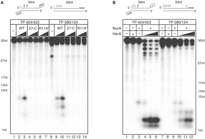
Figure 1.
Biochemical properties of PfNurA and PfHerA (A) Comparison of nuclease activities of PfNurA and dimeric interface mutants. TP 424/423 (lanes 1–7) or TP 580/124 (lanes 8–14) DNA substrates (20 nM) were incubated for 120 min at 65°C with increasing amounts of PfNurA (350 and 1750 nM) and 5 mM MnCl2. 27-C (Δ1–26) and R11A/I12E/S60Y (R11A*) are the PfNurA dimeric interface mutant proteins. Reaction mixtures were analyzed on 15% denaturing polyacrylamide gels containing 7 M urea in TBE buffer. Schematic diagrams of each substrate with a 32P-labeled 5′-end (asterisk) and cleavage sites (arrows) are shown on the top. Five nucleotides in one 3′-end are connected through phosphorothioate bonds, which are shown as ‘SSSSS’. ssDNA markers are indicated. (B) Nuclease activities of PfNurA in the presence of PfHerA. DNA substrates (20 nM) were incubated for 120 min at 65°C with 350 nM of PfNurA, increasing amounts of PfHerA (35, 70 and 115 nM), 5 mM MnCl2 and 1 mM ATP. Lanes 2 and 8 contain 115 nM PfHerA.

