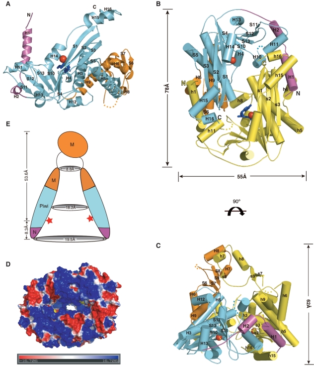
Figure 2.
Overall structure of Pf NurA. (A) Structure of the Pf NurA monomer. The N-(magenta), PIWI (aqua), and M (orange) domains are shown with Mn2+ ions (red spheres). (B) The flat side of the Pf NurA dimer is viewed from the front and (C) from a 90° rotated view (along the horizontal axis of Figure 2B). Three domains of one monomer are shown in the same color scheme as in 2A (magenta, aqua and orange) and another monomer is shown in yellow for clarity. Secondary structures are labeled. Red spheres represent Mn2+ ions, and the two dAMP molecules are colored in blue. The disordered region is shown as dots. Dimension of the central hole is 29 × 19 Å2. (D) Electrostatic potential of the Pf NurA dimer mapped onto the solvent-accessible protein surface (blue indicates positive regions; red indicates negative regions; the metal ion is in yellow). The figure is shown as a 90° rotated view of 2B along a 2-fold axis. (E) A schematic drawing of the Pf NurA central channel in the same view as that of 2C.The Mn2+ ions are represented as red stars.

