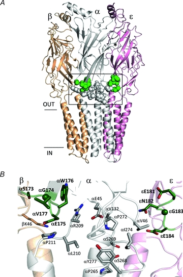
Figure 1. Location of the C-terminus of loop 9 of AChRs.

A, side view of the Torpedo AChR (accession number 2bg9.pdb). The mutated loop 9 positions in the α and ε subunits are shown as dark grey spheres (green spheres online); the residues tested for gating interactions with the loop 9 residues are shown as white spheres. *marks approximately the αε transmitter binding site and the horizontal lines mark approximately the membrane. The M4 transmembrane helices have been removed for clarity. B, higher resolution view of the boxed area in A. The residues are labelled according to their numbers and side chains in mouse AChRs (loop 9 residues, dark grey (green online) and bold).
