
Figure 3. Differentiation between pre- and postsynaptic LTPi.
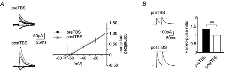
A, example traces of GABAA-receptor-mediated IPSCs at holding potentials of −5, −20, −40, −60 and −70 mV before and after TBS (preTBS and postTBS; left). Plot of normalized amplitudes vs. holding potential. The reversal potential of GABAA-receptor-mediated currents has been calculated by a linear fit of the current–voltage relationship (right). No significant shift of the reversal potential was observed after TBS (postTBS) as compared to baseline (preTBS). B, example traces (left) and quantification (right) of the PPR (PPR = amplitude2/amplitude1) at a paired-pulse interval of 50 ms. The LTPi induction by TBS significantly reduced the PPR as compared to the PPR during baseline conditions, indicating an increase of the release property at GABAergic presynaptic terminals (**P < 0.01; t test).