
Figure 4. nNOS is present in principal neurons of the BLA.
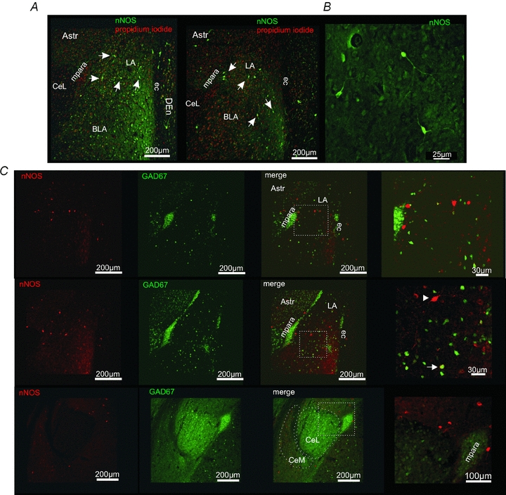
A, nNOS immunoreactivity in the amygdala. nNOS-positive (green) neurons and fibres could be detected in the BLA. Outside the amygdala nNOS-positive neurons were detected in the dorsal endopiriform cortex (DEn) close to the external capsule (ec), and the amygdalar/striatal transition zone (AStr). In the medial paracapsular interneuron clusters (mpara) no nNOS-positive somata could be detected. Propidium iodide (red) was used to stain cell nuclei. B, examples of nNOS-positive neurons within the LA. nNOS-immunoreactivity is visible in the soma and neurites of these neurons. C, immunohistochemical stainings for nNOS (red) in transgene GAD67-EGFP (green) mice. Although in a small subset of neurons a nNOS-GAD67-EGFP co-localization was detected (arrow), most of the nNOS-positive neurons were GAD67-EGFP negative in the BLA (arrowhead). The lateral region of the central amygdalar nucleus (CeL) is virtually devoid of nNOS-positive fibres.