
Figure 1. Concentration-dependent inhibition of GABAARs by TBPS and picrotoxin.
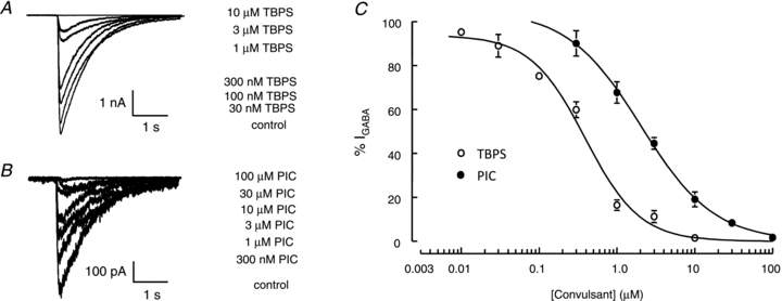
A and B, representative whole-cell currents recorded from α1β2γ2 receptors expressed in HEK293 membranes were activated by GABA (100 μm for 100 ms every 60 s) and inhibited by TBPS (A) and picrotoxin (B). C, concentration–inhibition curves for picrotoxin (•) and TBPS (○). GABA-evoked current amplitudes recorded in the presence of picrotoxin (n≥ 3) or TBPS (n≥ 4) are expressed as a percentage of those recorded under control conditions (%IGABA). TBPS caused a more potent inhibition of GABA-evoked currents. IC50 values for TBPS and picrotoxin were 0.39 ± 0.07 μm and 2.0 ± 0.2 μm, respectively. Data are represented as means ± SEM of 3–10 recordings.