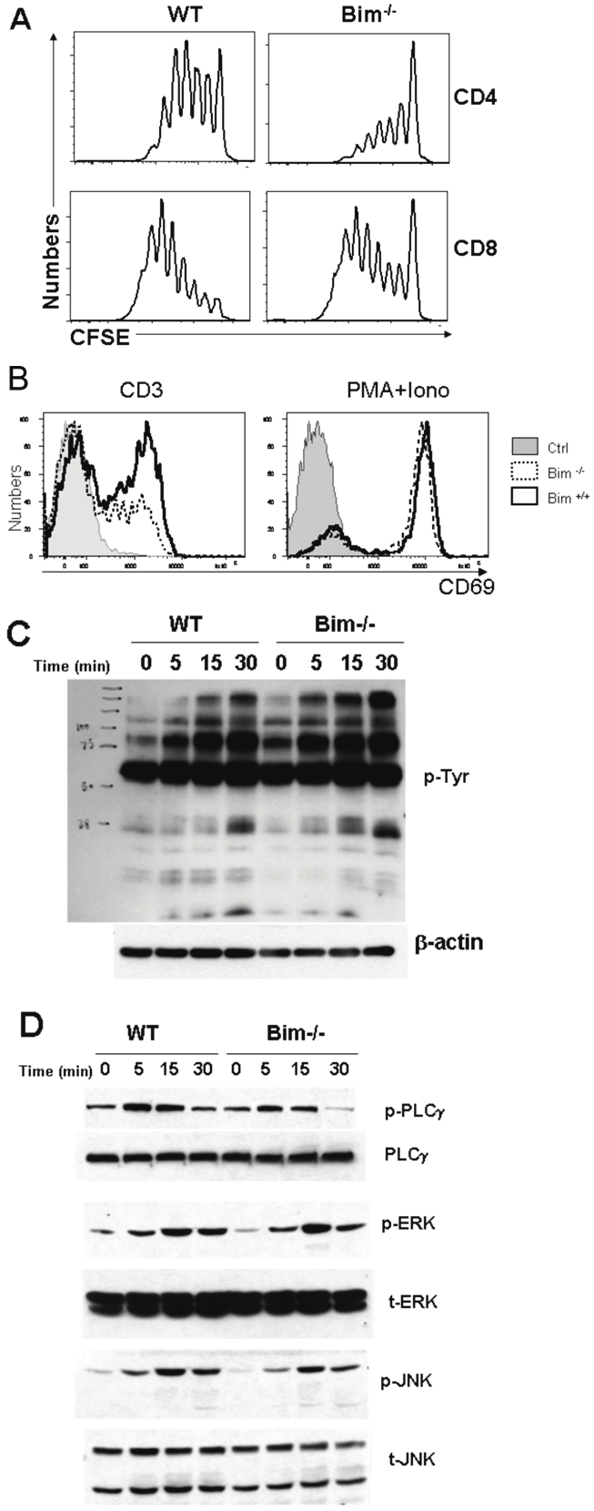
Figure 5.

Bim deficiency down-regulates TCR-induced CD69 expression. (A) T cell proliferation after anti-CD3 stimulation. Splenocytes from WT or Bim-/- B6 mice were labeled with CFSE and stimulated with anti-CD3 Ab for 3 days. CFSE dilution was measured by FACS analysis on CD4+ (top panel) and CD8+ cells (bottom panel). (B) CD69 expression. T cells from WT or Bim-/- B6 mice were cultured with medium alone (Ctrl), or stimulated by anti-CD3 mAb (left) or PMA + ionomycin (right) for 20 hrs. Cells were harvested and stained for CD69 expression, and the data represent 1 of 3 replicate experiments. (C) Total protein tyrosine phosphorylation was measured on the T cells from WT and Bim-/- mice stimulated with or without anti-CD3 mAb at 10μg/ml for 5, 15 and 30 min. (D) Tyrosine phosphorylation of PLCγ, ERK and JNK were analyzed by western-blot. Representative data from more than 3 similar experiments are shown.
