
Figure 1.
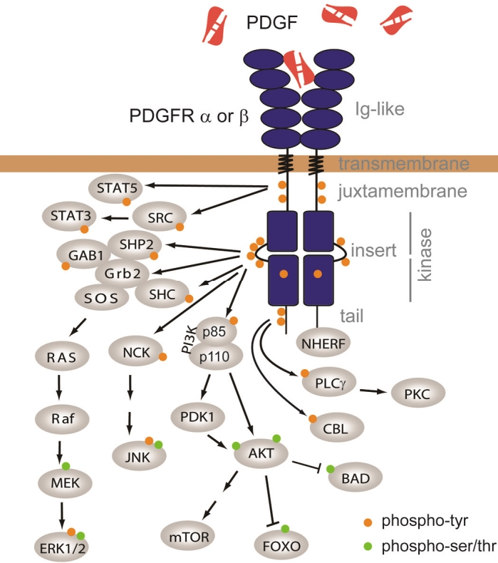
PDGF receptors and signaling. PDGFR domains are named in gray on the right. Arrows depict protein interaction and/or phosphorylation. Phosphorylation of tyrosines is represented by a orange disk, while phosphorylated serines and threonines are represented in green. See text for details.