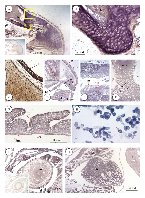
Figure 2.

ISLB staining of snout, alimentary canal, erythrocytes, ovary, and heart. (A), Snout; STC-1 binding sites in the snout are most prominent in cranial cartilage (c), the inset at the lower left is a staining control; (b), brain olfactory bulb; skm, skeletal muscle. (B), higher magnification of smaller boxed area in (A) showing intense binding activity confined to the matrix surrounding lacuna-bound chondrocytes (*). (C), tissue section adjacent to larger boxed area in (A), stained by ICC for STC-1 protein. In cranial cartilage (c), STC-1 is present in both chondrocytes and the surrounding matrix. High immunoreactivity is also present in skin (s). (D), Buccal cavity; binding sites are most evident in the mucosal (m) layer of cells lining the mouth and emerging teeth (*), the boxed area shows a magnified tooth (*) with high binding activity (red arrows) in the dentine layer (bc, buccal cavity; s, skin; p, pigment layer). (E), esophagus; weak binding is present in the mucosal (m) and sub-mucosal (sm) layers of the esophagus. Muscularis mucosa (mm) exhibits the highest binding activity. (F), stomach; binding is lowest in mucosa (m) and highest in the underlying muscularis mucosa (mm) at the junction of the cardiac (c) and pyloric (c) regions of the stomach. (G), small intestine; binding is highest in mucosal enterocytes (m) and muscularis mucosa (mm) and lowest in the submucosa (sm). (H), red blood cells; most erythrocytes contain high binding activity that is confined to the cytoplasm. (I), ovary, the highest binding is confined to the cytoplasm of primary oocytes (po). Weaker binding is evident in theca cell (tl), granulosa cell layers (gl), and cytoplasm of vitellogenic oocytes (vo). The inset at the lower left is a staining control. (J), heart (h); all cardiac myocytes exhibited high binding activity as did surrounding skeletal myocytes (skm). Lower binding was present in liver (l) hepatocytes.
