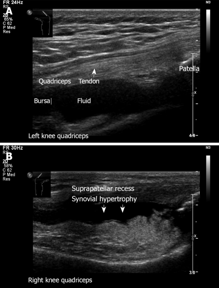
Figure 16.
Ultrasound examination of knee quadriceps. A: Left knee longitudinal view. Transducer on the anterior aspect of the knee. Note the fluid in the suprapatellar recess. Normal quadriceps tendon with its insertion to the patella; B: Similar transducer position demonstrating synovial hypertrophy. A 68-year old woman with rheumatoid arthritis presented with pain and swelling of the right anterior knee.

