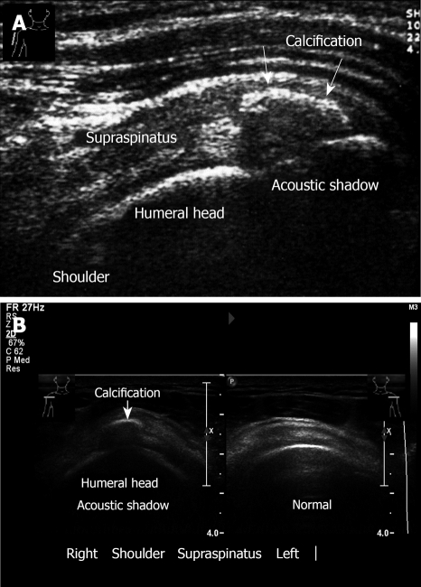
Figure 6.
Ultrasound examination of calcification. A: Calcific tendinitis. Calcification in the supraspinatus tendon, longitudinal view. Note: Acoustic shadow behind the calcification. A 29-year old woman presents with a short history (3 d) of incapacitating shoulder pain and with severe restriction in the range of shoulder movement; B: Transverse sonogram. Calcification of supraspinatus, right. Left normal sonogram.

