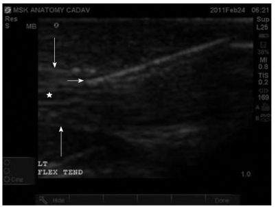
Figure 5.
Tendon injection can be performed in either transverse or longitudinal planes. Insertion of the needle in long-axis allows excellent visualization of the tendon sheath and tendon fibers. Prior to injection the needle tip should be well-visualized between the tendon sheath and the tendon fibers. An important method to ensure needle visualization is to angle the probe to make the angle of ultrasound wave as close to perpendicular as possible. Additionally, confirmation of a structure as a tendon involves angling the probe along the tendon to note tendon anisotropy characteristic of a tendon and not noted in nerves or vessels. Short arrow indicates needle tip in long-axis section. Long arrows indicate tendon sheath. Star indicates tendon fibers in long-axis section.

