
Figure 7.
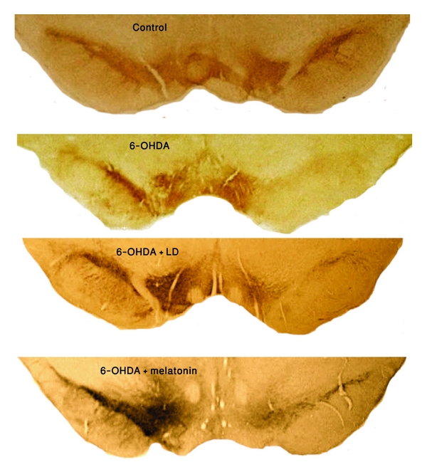
Representative TH-immunostained from coronal sections containing the SNc of control, 6-OHDA-lesioned, 6-OHDA-lesioned + LD and 6-OHDA-lesioned + melatonin-treated rats. Note the profound cell loss in the ipsilateral SNc in the three experimental groups, being more evident in the 6-OHDA and LD treated ones; also, the contralateral SNc of melatonin-treated rats lost fewer neurons than the other two experimental groups (magnification 4×).