
Figure 8.
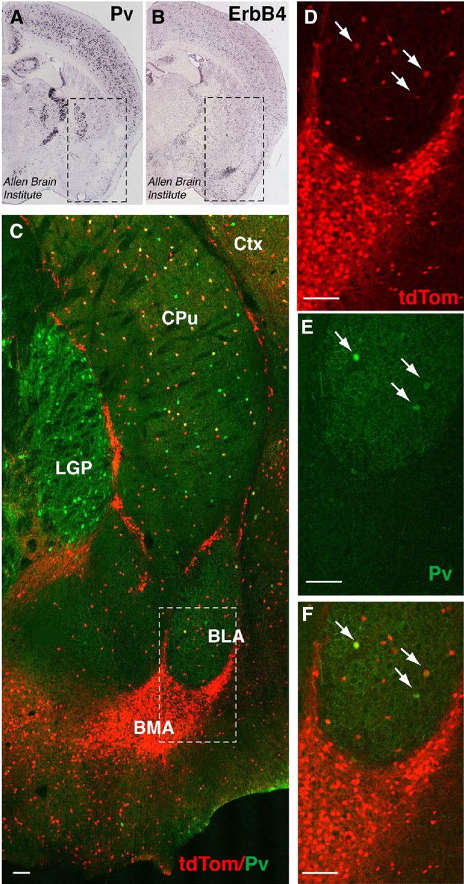
ErbB4 is abundantly expressed in the amygdala and shows only modest colocalization with PV. A, B, In situ hybridization of PV and ErbB4 in coronal sections of P56 mice (Allen Brain Atlas, 2009). Boxed areas correspond to the region shown in C and include the amygdala (ventrally), caudate/putamen, and the lateral global pallidus (dorsally). PV mRNA signals are dense in the lateral globus pallidus (LGP), sparse in the BLA, and not detectable in the BMA. By contrast, ErbB4 mRNA signals are very dense in the BMA and more sparsely distributed in most other areas. C, Co(immuno)fluorescence micrograph image of tdTomato (tdTom; red), reporting ErbB4 expression in adult Ai14 × ErbB4-2A-CreERT2 mice, and endogenous PV (green). The boxed area is magnified in D–F. While few tdTomato-labeled cells in the BLA coexpress PV (arrows), virtually none of the large number of the ErbB4-expressing cells in the BMA and other intercalated areas coexpress PV. CPu, Caudate–putamen; Ctx, cerebral cortex; LGP, lateral globus pallidus. Scale bar, 100 μm.